
Clinician’s Corner – Educational Content for Clinical Directors,
Chief Dental Officers, Clinical Boards and the Entire Clinical Team.
Case Study by Troy Schmedding, DDS, AAACD. Sponsored by Garrison Dental
Composite resins are, and for the foreseeable future, the most widely used direct restorative material for restorations involving anterior teeth. Today’s composite materials offer practitioners tremendous physical properties but also optical properties to deliver lifelike restorations on a consistent basis. Proper finishing and polishing, together with the right occlusal scheme can provide the opportunity for these restorations to last a long period of time.
Though the handling characteristics of modern composites are vastly improved dentists still struggle with the development of good inter-proximal contacts and proper anatomical contours. Utilizing an anterior matrix system can be paramount in achieving this objective. Matrix systems in the anterior region are numerous in respect to creativity but the majority can be classified into flexible and rigid. Flexible matrices include the popular mylar strip and soft splint templates that can be challenging as they lack the ability to contour large areas leading to irregular contours and contacts. Rigid matrices include putty indexes and pre-contoured posterior sectional matrix, both viable options in certain situations but the majority of time posterior matrices are cumbersome and difficult to use when restoring multiple surfaces.
To overcome a majority of these aforementioned limitations Garrison Dental has introduced the Fusion Anterior Matrix System. A simple matrix designed to be used for anterior restorations such as class III, IV as well as composite veneers. The firm stain-less steel matrix easily slides into the sulcus all while maintaining the proper shape and contour without deformation. Properly placed the ideal anatomical curvature is achieved in a gingival – incisal and facial – lingual direction. To help maintain this ideal anatomical position the Fusion Anterior Wedge is used to ensure a firm seal at the cervical margin from facial to lingual. These radically curved wedges help free up your hands to allow one to concentrate on composite placement and simplify the restorative process.
This article is a case report of a direct composite restoration on a maxillary anterior tooth where the proximal contact and incised edge position were developed using the Fusion Anterior Matrix System.
Case Report:
A 74 year old male presented with an old class III composite on the distal portion of his left lateral incisor (Figure 1). Re-current decay was noted both visually as well as radiographically. Treatment options were discussed with patient and it was decided we would replace the restoration with a new direct composite restoration. Small amounts of composite were placed on to the tooth and light cured to get an idea of what shade or shades would be utilized.
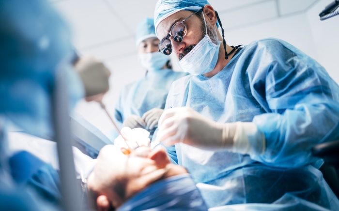
The patient was anesthetized with 1/2 carpule of 4% Articaine (Septodont) with 1:100,000 epinephrine. Isolation was obtained with a ComfortView® Lip & Cheek Retractor (Premier Dental). A pear shaped diamond bur (Meisinger) was used to re-move the old restoration. A #2 round bur in a slow speed handpiece was used to re-move all remaining decay and finally an 856 diamond bur (Meisinger) was used to bevel the facial margin and create a butt margin on the lingual.
A short anterior Fusion Band (Garrison Dental) was then placed inter proximal past the finish line of the preparation and gently into the sulcus. A medium sized anterior Fusion Wedge (Garrison Dental) with its radical curvature was placed to ensure a firm seal at the cervical margin all while not distorting the anatomical contour, unlike the mesial aspect of this same tooth where a wood wedge was used to seal the margin of that particular restoration (Figure 1). A smaller wedge may have been an option in this case as you can you see the orange wedge rotated slightly when placed but due to the lack of distortion of the band and the great seal achieved I did not change.
Access to both the facial and lingual of the preparation achieved a 35% phosphoric acid (K-Etchant Kuraray) was placed in a selective etch protocol and allowed to sit for 30 seconds prior to rinsing off (Figure 2). Universal Bond Quick (Kuraray) was then applied to the preparation in agitating motion for 10 seconds, air thinned and light cured for 20 seconds from both the facial and lingual to ensure polymerization. In the case of using a metal matrix that doesn’t promote light passage as friendly as a clear matrix I felt more comfortable taking the extra time to polymerize. The flexibility of the Fusion Matrix system allows you to sculpt composite in an open fashion where you have access from the lingual as well as the facial while still benefiting from the anatomically correct inter-proximal contours.
In this particular case I took the approach of using my finger to push the matrix from the facial while adding a small increment of Majesty ES-2 Universal composite (Kuraray) from the lingual (Figure 3). Composite instrument was then used from the lingual to manipulate the composite (Figure 4) and then light cured for 20 seconds. Repeated this process (Figure 5) until the restoration was filled to completion and light curing finished from the lingual. The matrix was then peeled away on the facial side and again cured for 20 more seconds to ensure complete polymerization. Anterior Fusion Wedge and Matrix were then removed to show minimal excess composite on the facial surface as well as ideal anatomical contours (Figure 6). A fine flame diamond and a fine football shaped diamond (Meisinger) did most of the heavy refinement and adjustment of occlusion. The final polish was achieved using a two step diamond polishing system (Meisinger)(Figure 7).

(Figure 1) Failing Class III composite on the distal – facial of the Maxillary lateral incisor

(Figure 2) Fusion Anterior Matrix in place with Fusion Wedge to stabilize matrix and allow easy access for placement of phosphoric acid .

(Figure 3) Fusion Matrix being held from the facial while placing composite from the lingual.

(Figure 4) Instrumentation of the composite performed to push the composite against the facial side of the Fusion Matrix

(Figure 5) Application of composite continues from the lingual til preparation is sufficiently filled.

(Figure 6) Removal of the matrix reveals proper contact, contours and minimal flash on the facial surface.

(Figure 7) Final polished restoration with wonderful aesthetics and ideal anatomical form and function.
Understanding the limitations of the material in every situation and adapting newer techniques should be our focus to make our restorations more predictable and durable. The case presented featured a great new product that can help you establish good contacts and contours in the anterior region where paramount importance is not just from an aesthetic but also a functional point of view.
For more information on the featured product click HERE.

To view instructional videos on this and more from Garrison Dental visit their YouTube channel HERE.
REFERENCES:
1) Ayush Goyal, Vineeta Nikhil, and Ritu Singh, “Diastema Closure in Anterior Teeth Using a Posterior Matrix,” Case Reports in Dentistry, vol. 2016, Article ID 2538526, 6 pages, 2016.
2) Fellippe L. A., Monteiro S. Jr, De Andrada C. A., Di Cerqueira A. D., and Ritter A. V.. 2005. Clinical strategies for success in proximo-incisal composite restorations. Part II. Composite application technique. J. Esthet. Restor. Dent. 17:11–21.
3) 2015 Oct;31(10):1214-24. doi: 10.1016/j.dental.2015.07.005. Epub 201Aug21.Anterior composite restorations: A systematic review on long-term survival and reasons for failure
Flávio F Demarco 1 , Kauê Collares 2 , Fabio H Coelho-de-Souza 3 , Marcos B Correa 2 , Maximiliano S Cenci 2 , Rafael R Moraes 2 , Niek J M Opdam 4
4) A systematic approach to contouring and polishing anterior resin composite restorations: A checklist manifesto. Vargas MA, Margeas R.
J Esthet Restor Dent. 2021 Jan;33(1):20-26. doi: 10.1111/jerd.12698. Epub 2020 Dec 25.
 Case Study by Troy Schmedding, DDS, AAACD. Dr. Troy Schmedding is a 1993 honors graduate of the Arthur A. Dugoni School of Dentistry in San Francisco, California. He maintains a private practice in Walnut Creek, Ca. where he focuses on aesthetic and functional dentistry. An Accredited member of the American Academy of Cosmetic Dentistry, he lectures both nationally and internationally on aesthetics and restorative materials. He has also written and published numerous articles on restorative materials and protocols in numerous dental magazines. Dr Schmedding also serves as a Key Opinion Leader for numerous manufacturers helping develop and bring new products to market. He can be reached at Troyschmeddingdds@gmail.com