
Clinician’s Corner – Educational Content for Clinical Directors,
Chief Dental Officers, Clinical Boards and the Entire Clinical Team.
Written by Dr. David Landwehr, Capital Endodontics
Sponsored by Surround Medical Systems
What is Stationary Intraoral Tomosynthesis (sIOT)?
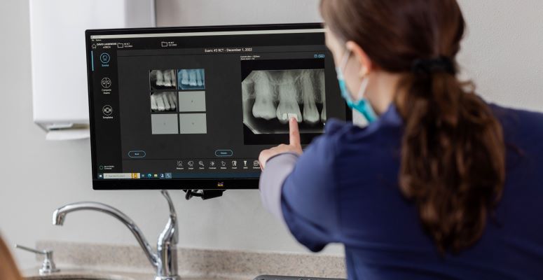
Stationary Intraoral Tomosynthesis is a disruptive, cutting-edge imaging technology being used by Surround Medical Systems in their Portray® intraoral imaging systems that is intended to change the way dentists use radiographs in the diagnostic process. The Portray system uses a carbon nanotube array to capture multiple images with a single exposure from multiple sources (Figure 1 black arrow). This generates a series of images that are then manipulated by a computer algorithm, like cone beam computerized tomography (CBCT), to generate an image stack that allows visualization of the field through or around the teeth. Each reconstruction stack contains buccal to lingual slices, with each slice representing 0.5 mm of object thickness. Each reconstruction also allows rotation of a 2D image plus or minus 6 degrees to open interproximal contacts. The Portray system is also able to take a standard 2D image should the need arise however with the introduction of sIOT the need for standard 2D images will quickly become obsolete.
The image sensor is coupled to the tube head with four magnets. This standardizes the x-ray source to sensor distance which allows the software to determine buccal-lingual distances. As a result, it is possible to visualize palatal roots of maxillary molars without superimposition of the sinus or buccal roots – resulting in better lesion detection at the palatal root when considering treatment. A single sIOT image provides more data and more information translates into more diagnostic confidence for dentists.
Does sIOT impact clinical workflow?
Image acquisition is virtually identical to periapical imaging because it is a standard size 2 digital sensor. Sensor placement is the same as for traditional two-dimensional periapical images. As a result, limited training was necessary for my dental assistants to acquire the images. The time required to obtain the image is slightly longer for sIOT compared to periapical images with a seven second acquisition time. However, because the tube is indirectly attached to the sensor it eliminates the need for retakes if the film placement is accurate and there is no motion blur to the images because there is no movement of the x-ray tube. Additionally, the acquisition time is likely to come down as the speed of the sensor increases and it is interesting to note that the sensor is already the fastest intraoral digital sensor used in dentistry.
After the image is taken the computer immediately creates the stack of images and there is no lag time following exposure of the image before I can manipulate the images and begin the diagnostic evaluation. The image resolution is identical to standard two-dimensional digital imaging. The viewing software, although powerful, is intuitive to use from day one and I did not need any advanced training to review the image stack. As a result, my workflow is identical compared to conventional two-dimension imaging, but more information is obtained resulting in better diagnosis and a better patient experience with less retakes.
What are the advantages and disadvantages of sIOT compared to conventional imaging and CBCT?
As previously stated, sIOT provides more diagnostic information than conventional periapical imaging with no disruption in workflow. The only measurable disadvantage of sIOT compared to periapical imaging was my initial cost of changing systems. However, with increased caries detection, periapical lesion identification and periodontal diagnosis the return on investment is significant and cost should not be considered a barrier for clinicians who want to practice at the absolute highest level.
SIOT is not meant to replace CBCT. The amount of information on a sIOT compares favorably to periapical imaging, but more information is available on a CBCT, especially when considering the axial view that is available on a CBCT. However, this is not without significant concerns for many clinicians. Even with a small field CBCT a high number of incidental findings are likely to be present on a CBCT. Clinicians are responsible for all information visible on the CBCT and any subtle pathology that is not identified exposes the dentist to significant liability. As a result, a complete and thorough reading of the entire volume will either require a disruption in workflow throughout the day or a lot of time at the end of the day reviewing scans. Additionally, obtaining high quality, diagnostic scans require training of the staff while reading the scans requires special training for clinicians. Another disadvantage of CBCT relates to image acquisition and movement of the radiation source. The long acquisition time and movement of the CBCT source around the patient can result in metal artifacts and a noisy non-diagnostic image depending on the presence of metal restorations, implants, and patient movement.

How do radiation dosages compare between conventional periapical images, CBCT and sIOT?
Despite the longer exposure time compared to a standard periapical image the radiation dosage is of negligible difference compared to standard imaging because most of the seven second acquisition time is reserved for the sensor to reset itself. For example, a standard periapical image has a radiation dosage of 9.5 uSV while a single sIOT image has approximately 17 uSV. A standard 2D image using the Portray system has only 4uSV of radiation. This compares to a full mouth series of images that would be approximately 85 uSV. CBCT would have a range of dosages from a small field CBCT with 84 uSV to a large field of view with a mean dosage of 212 uSV. Although the uSV numbers reveal relative dosages, for patients it might be better to describe the radiation in terms of background radiation in days. For example, a standard periapical image is comparable to one day of background radiation, while a sIOT image is roughly 1.5 days of background radiation. A full mouth series of images or a small field CBCT have similar dosages approximating ten days of background radiation.
What are the clinical applications of sIOT?
The technology is so new at this point that clinical studies have not yet verified how much better sIOT will prove to be compared to conventional periapical imaging. However, the ability to rotate the sIOT image has proven to be far superior in my clinical practice for interproximal and recurrent caries detection compared to standard periapical imaging because of the opportunity for improved visualization of the contacts. Periapical lesion detection is also much improved compared to conventional imaging because of the ability to slice through the image stack in the buccal-lingual direction. Additionally, the ability to slice through the image stacks in a buccal-lingual direction allows for better evaluation of crestal bone height in the detection of periodontal disease. Currently there is no imaging technology that can readily show vertical root fractures and cracks. The reduction in metal artifacts and the ability to slice through and rotate around teeth with sIOT may result in increased crack detection as well as earlier identification of resorptive defects.
What does the future hold for sIOT?
The technology is very new, and most clinicians don’t know it exists or what the potential of sIOT might be. However, sIOT is being taught at a handful of dental schools in the United States and this should result in clinically relevant studies confirming its value and benefit compared to other imaging modalities. Software and hardware upgrades are likely to yield even better and quicker image acquisition and quality. It is possible, in the future, the unit will be on small cart allowing clinicians the freedom to more cost effectively obtain sIOT images in all operatories. Lastly, the image analysis will likely be coupled with artificial intelligence tools allowing practitioners to evaluate the images stacks more quickly with increased diagnostic accuracy. 
